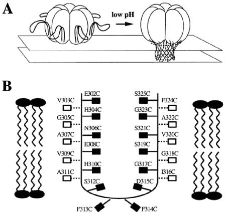
FIG. 1.
Model of acidification-induced pore formation by PA63. (A) After the pH is lowered, the 2β2-2β3 loops move to the base of the heptamer and combine to form a 14-stranded transmembrane β-barrel. (B) Hypothetical orientation of the 2β2-2β3 loop in the membrane, showing the sites of cysteine substitutions. This figure is adapted from Fig. 1 and 6 in Benson et al. (4). The filled boxes indicate residues that are expected to be in contact with the hydrophobic core of the membrane, while the open boxes represent residues that are expected to face the lumen of the pore.

