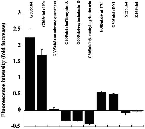
FIG. 5.
Effects of various reagents on the increase in fluorescence of NBD-labeled PA G305C. Cells were preincubated with 10 mM β-methyl-cyclodextrin for 1 h, 50 mM cytochalasin D for 35 min, or 1 μM bafilomycin A1 for 35 min at 37°C. For the experiment involving membrane quenchers, cells were preincubated with an equimolar mixture of 5-doxyl and 12-doxyl stearic acid for 1 h at room temperature. For the DNI experiment, NBD-labeled PA G305C was mixed with an equivalent amount of DNI before addition to cells. After each pretreatment, cells were incubated on ice for 1 h with the NBD-labeled PA. After washing, the cells were monitored for fluorescence intensity as described in Fig. 2. In one sample, LFN was added to a final concentration of 5 μM immediately after the sample was placed in the fluorimeter. Each bar represents the ratio of fluorescence at 1 h to that at time zero. NBD-labeled K563, a residue on the cap of the PA heptamer, was included as a control. Error bars represent standard deviations.

