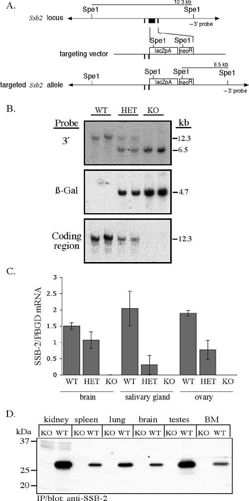
FIG. 2.
Disruption of Ssb-2 by homologous recombination. (A) The exons for Ssb-2 are shown in black (top). The targeting vector (middle) replaces the entire coding region with the β-galactosidase PGKneo cassette (bottom). (B) Southern blot analysis of SpeI-digested genomic DNA from the tails of mice derived from a cross between SSB-2+/− mice. The blot was hybridized with the 3′ Ssb-2 genomic probe, which distinguishes between endogenous (12.3-kb) and targeted (6.5-kb) alleles (top panel). The blot was then hybridized with a 266-bp probe corresponding to the coding region for β-galactosidase (center panel) and an 833-bp probe corresponding to the coding region for SSB-2 (bottom panel). (C) Expression of SSB-2 mRNA as measured by Q-PCR in tissues of wild-type (WT), SSB-2+/− (HET), and SSB-2−/−(KO) mice. SSB2 mRNA levels were normalized against PBGD mRNA levels. All bars represent averages and standard deviations for samples derived from three mice. (D) Lysates (1 mg of protein, except for bone marrow [BM; 0.36 mg]) from a variety of wild-type and SSB-2−/− tissues were analyzed by immunoprecipitation (IP) with anti-SSB-2 antibodies and Western blotting with biotinylated SSB-2 antibodies.

