Figure 3.
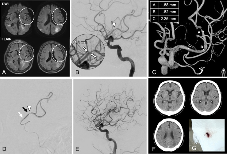
A 74-year-old man was hospitalized with a minor stroke, displaying new-onset right-sided paralysis, aphasia, and a National Institutes of Health Stroke Scale score of 16. (a) The MRI did not detect any evidence of acute cerebral infarction in the occluded vessel area (circle). (b) The lateral angiogram demonstrated M2 occlusion (arrowhead). (c) The occluded vessel diameter was 1.8 mm (white arrow). The treatable tortuosity and pathway were revealed using 3D rotational angiography. (d) A 1.7-mm outer diameter aspiration catheter established contact with a clot (arrowhead) via the solo contact method. Tip of the aspiration catheter (black arrow) and that of the microcatheter (white arrow), which was pulled proximally. (e) Complete recanalization visualized by angiography. (f) Lack of hemorrhage on CT. (g) Retrieved clot.

