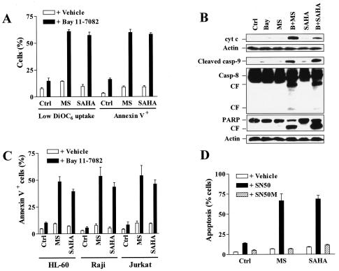
FIG. 4.
NF-κB inhibitors potentiate mitochondrial dysfunction and apoptosis mediated by HDAC inhibitors. (A) U937 cells were exposed for 24 h to 2 μM MS-275 (MS) or 1 μM SAHA in the absence or presence of 3 μM Bay 11-7082, after which the percentages of cells exhibiting decreased ΔΨm, reflected by low DiOC6 uptake, and annexin V positivity were determined by flow cytometry as described in Materials and Methods. (B) Alternatively, a Western blot analysis was performed to assess expression of cytosolic cytochrome c (cyt c) and cleavage of caspases and PARP. CF, cleavage fragment; Casp, caspase; Ctrl, control; Bay, Bay 11-7082; MS, MS-275; B+MS, Bay 11-7082 plus MS-275; B+SAHA, Bay 11-7082 plus SAHA. Each lane was loaded with 30 μg of protein; blots were subsequently stripped and reprobed with anti-β-actin to ensure equivalent loading and transfer of protein. The results are representative of three separate experiments. (C) HL-60, Jurkat, and Raji cells were exposed to 2 μM MS-275 (MS) or 1 μM SAHA for 24 h in the absence or presence of Bay 11-7082 (HL-60, 3 μM; Jurkat, 2 μM; Raji, 5 μM), after which the percentage of annexin V-positive cells was determined. (D) U937 cells were incubated for 24 h with 2 μM MS-275 (MS) or 1 μM SAHA in the absence and presence of 150 ng/ml NF-κB inhibitory peptide (SN50) or control negative peptide (SN50M), respectively, after which the percentage of cells exhibiting apoptotic morphology was determined as described in Materials and Methods. For panels A, C, and D, results represent the means ± SD for three separate experiments performed in triplicate.

