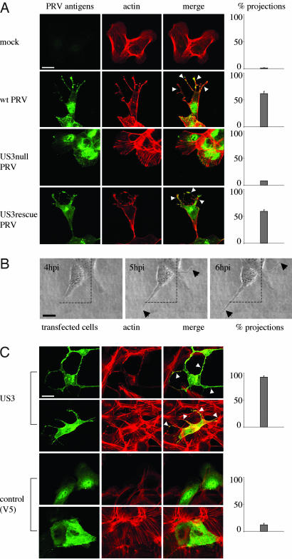
Fig. 1.
US3 mediates the formation of long, actin-containing cell projections in PRV-infected and US3-transfected cells. (A) ST cells that were seeded at low density (cell clusters of 5–10 cells) were either mock-infected or infected with wild-type, US3-null, or US3-rescue PRV at a MOI of 10. Cells were paraformaldehyde-fixed at 7 hpi, permeabilized, and stained for viral proteins (green) and actin (red). Arrowheads indicate cell projections. (Bar, 10 μm.) (Right) Graphs showing percentages of cell clusters that display projections (means ± SD of triplicate assays). (B) Live cell imaging of the formation of cell projections in PRV-infected cells. ST cells were inoculated with PRV at a MOI of 10 and analyzed at different time intervals by using phase contrast microscopy. Arrows indicate cell projections. (Bar, 10 μm.) (C) ST cells were transfected with a eukaryotic expression vector encoding the PRV US3 protein (Upper) or a control protein (V5, Lower). At 24 h after transfection, cells were paraformaldehyde-fixed, permeabilized, and stained for US3 or V5 (green) and actin (red). Arrowheads indicate cell projections. (Bar, 10 μm.) (Right) Graphs showing percentages of transfected cells that display projections (means ± SD of triplicate assays).

