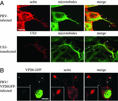
Fig. 2.
US3-induced cell projections contain microtubules in PRV-infected and US3-transfected cells and virus particles in PRV-infected cells. (A) ST cells, at 7 hpi with wild-type PRV (Upper) or 24 h after transfection with a US3-encoding plasmid (Lower), were paraformaldehyde-fixed, permeabilized, and stained for microtubules (green) and actin (red, Upper) or US3 (red, Lower). Arrowheads indicate cell projections. (Bar, 10 μm.) (B) ST cells, at 7 hpi with a PRV strain that encodes a GFP-tagged VP26 capsid protein (green), were paraformaldehyde-fixed, permeabilized, and stained for actin (red). Small green dots represent virus particles. Regions with virus particles are indicated with arrowheads. (Bar, 10 μm.) (Inset) An enlargement of the boxed area.

