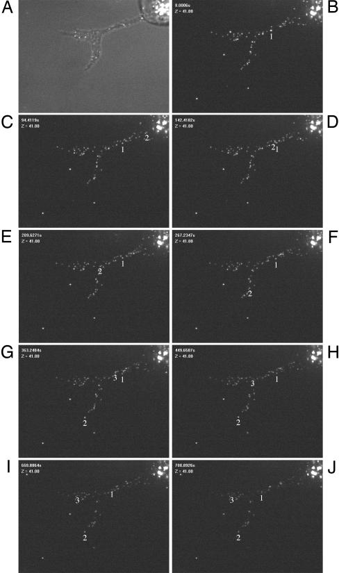
Fig. 3.
PRV capsids travel in the US3-induced cell projections. ST cells were infected at a MOI of 10 with a PRV strain that expresses a GFP-tagged VP26 capsid protein. At 6 hpi, a live cell imaging video was taken from a cell that displays a cellular projection (A, phase contrast image of the cell; B–J, time lapse recording images, total duration, 910 s, taken with 10-s intervals). Different capsid movements could be observed (see Movie 1). Individual movement of three capsids are summarized (tracking number is located underneath particle). Capsids 2 (indicated with an arrow in Movie 1) and 3 show fast, saltatory migration toward the tip of the projection. Capsid number 1 shows short migration toward the nucleus (B–D), which was only rarely observed, later followed by movement toward the tip of the projection (H–J). (Bar, 5 μm.)

