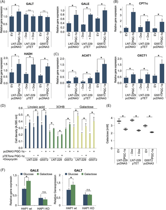
FIGURE 3.

Overexpression of PGC‐1α facilitates use of alternative nutrients. (A–C) cDNA of LNT‐229 and G55T2 EV and PGC‐1α cells and LNT‐229 pTetOne PGC‐1α cells with and without 0.1 µg/mL doxycycline (± Dox) cultured in serum‐free medium for 24 h was generated. Gene expression of GALT and GALE (A), CPT1c and HAHD (B), and OXCT1 and ACAT1 (C) was quantified; values are normalised to 18S as well as SDHA housekeeping gene expression (n = 3, mean ± SD, *p < 0.05, **p < 0.01). (D) The cells were exposed to serum‐free medium without glutamine containing either 25 mM galactose or 2 mM glucose with the addition of 100 µM linoleic acid or 5 mM 3OHB. Cell density was measured by crystal violet staining after 3 days (n = 3, mean ± SD). (E) Cells were incubated in medium containing 5 mM galactose for 6 h. Remaining galactose in the medium was determined (n = 4, mean ± SD, *p < 0.05). (F) cDNA of HAP1 wt and HAP1 PGC‐1α KO cells cultured in 25 mM glucose or 25 mM galactose for 5 days was generated. Gene expression of GALE and GALT was quantified (n = 3, mean ± SD, *p < 0.05).
