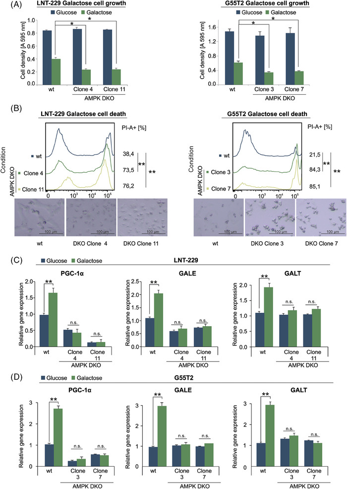
FIGURE 5.

Knockout of AMPK disturbs galactose metabolism. (A, B) LNT‐229 (left panel) and G55T2 (right panel) wt and AMPK DKO cells were incubated in medium containing 25 mM glucose or 25 mM galactose. Cell density was measured by crystal violet staining after 3 days (A). Cell death was determined by propidium iodide staining after 48 h (B). Representative photographs of the cells are included below the panels (bright field microscopy, 48× magnification). (C, D) cDNA of LNT‐229 (upper panel) and G55T2 (lower panel) wt and AMPK‐DKO cells maintained in culture medium with either glucose or galactose for 5 days was generated. Gene expression of PGC‐1α, GALE and GALT was quantified (n = 3, mean ± SD, *p < 0.05, **p < 0.01).
