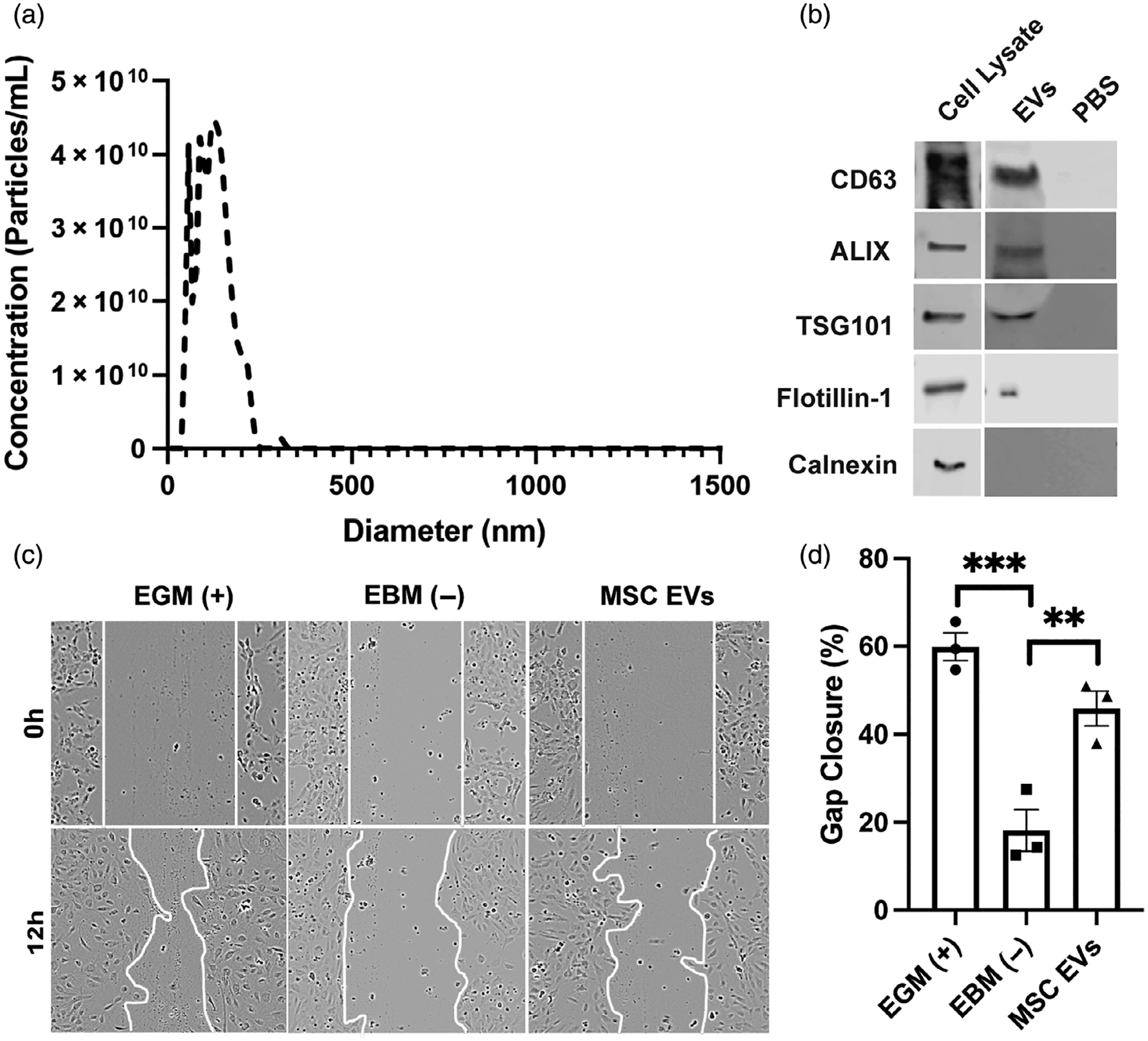
FIGURE 1.

Extracellular vesicle (EV) characterization and bioactivity assessment. (A) Nanotracking analysis of mesenchymal stem/stromal cell (MSC) EVs. (B) Immunoblots of EV-associated markers CD63, ALIX, TSG101, and Flotillin-1 as well as endoplasmic reticulum (ER) marker (Calnexin). (C, D) Human umbilical vein endothelial cells (HUVECs) were treated with growth media only (EGM; positive control), basal media only (EBM; negative control), 100 μg/ml MSC EVs in basal media. Images were taken at time 0 (brightfield) and 12 h post wounding of the cell layer. Representative images for each group and timepoint are displayed and the gap area outlined with a white line. Statistical significance was calculated using one-way ANOVA with Holm–Sidak’s test (**p < .01, ***p < .001) (n = 3)
