Fig. 1.
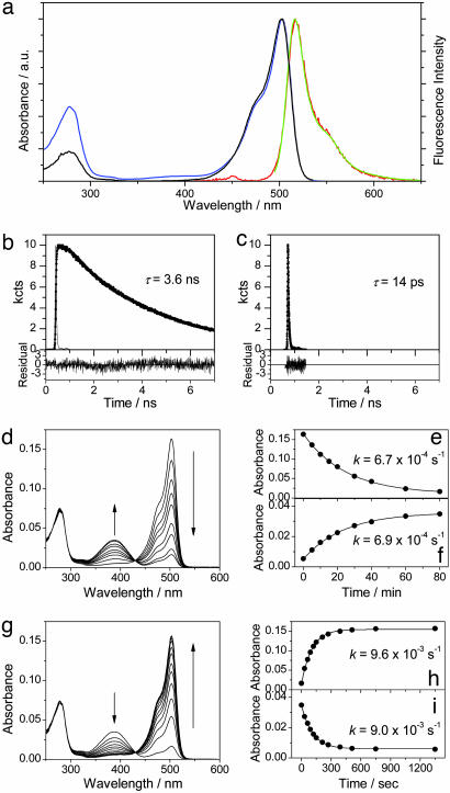
Photoswitching of Dronpa at the ensemble level. (a) Steady-state spectra of Dronpa at pH 7.4; absorption spectrum (blue line), fluorescence spectrum excited at 488 nm (green line) and 390 nm (red line), and excitation spectrum detected at 540 nm (black line). a.u., arbitrary units. (b and c) Fluorescence decays of Dronpa (pH 7.4) excited at 488 nm and detected at 520 nm (b) and excited at 390 nm and detected at 440 nm (c). (d) Time evolution of the absorption spectrum of Dronpa (pH 7.4) on irradiation with a 488-nm laser (P = 10 mW/cm2). (e and f) Time evolution of the peak absorbance of the deprotonated (e) and protonated (f) form on irradiation with a 488-nm laser. The solid lines show the fitting with a first-order kinetic model. (g) Time evolution of the absorption spectrum of the photoswitched Dronpa (488-nm irradiation) on irradiation with a 405-nm laser (P = 1 mW/cm2). (h and i) Time evolution of the peak absorbance of the deprotonated (h) and protonated (i) form on irradiation with a 405-nm laser. The solid lines show the fitting with a first-order kinetic model.

