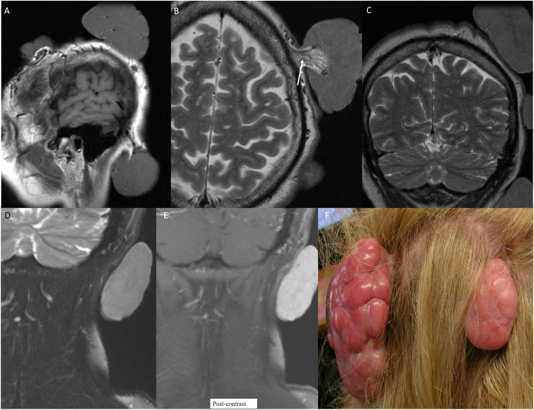
Figure 6.
Multiple variable sized scalp nodules in a 55-year-old with Brooke–Speigler Syndrome. Multiple MR images (a-e) reveal large scalp nodules including few sessile and few pedunculated. Pedunculated nodule along the left scalp shows highly vascular pedicle (b, arrow). Avid contrast enhancement within the lesion on coronal post-contrast image (e). Photograph of the lesions (f) depicting the large multilobulated nodules of the scalp. Biopsy of nodules revealed spiradenomas.

