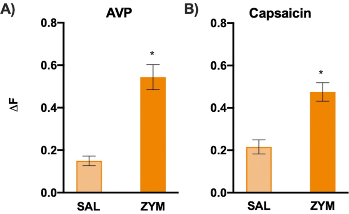
Figure 8.

In vitro Ca2+ imaging of ENS neurons from VH mice exhibits significantly greater Ca2+ influx to AVP and capsaicin. (A) In vitro Ca2+ imaging of ENS neurons revealed individual neurons from VH mice (BL/6NTac-ZYM) exhibited significantly greater Ca2+ influx (ΔF) in response to AVP compared with SAL controls (t = −4.556, P < .001). Refer to Supplementary Fig 8 for individual ΔF distributions for each condition and agonist. (B) ENS neurons revealed individual neurons from VH mice also exhibited greater Ca2+ influx (ΔF) in response to capsaicin compared with SAL controls (t = −4.838, P < .001). The % of neurons responding to AVP and capsaicin did not differ significantly (data not shown) (all P > .05). Refer to Supplementary Fig 9 for representative traces of enteric neuron responses for each condition and agonist. * Indicates significant t-test, P < .05.
