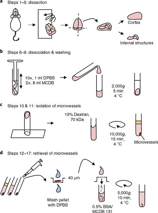
Fig. 1 |. Cortical microvessel isolation protocol overview.
a, Dissection. After mouse euthanasia, all steps are conducted in a cold room. Meninges are removed by rolling the brains on blotting paper, and cortices are dissected in cold Dulbecco’s phosphate-buffered saline (DPBS). b, Dissociation of tissue and washing. Cortices are homogenized using a loose-fit Dounce grinder and centrifuged (4 °C) for 5 min at 2,000g. c, Separation of the microvessel fraction by gradient centrifugation. Microvessel pellet is resuspended in a 15% (wt/vol) 70-kDa dextran solution and centrifuged (4 °C) for 15 min at 10,000g. d, Retrieval of microvessels. The top layer, containing myelin and brain parenchymal cells, is removed. Microvessels are retrieved by pipetting and transferred to a 40-μm cell strainer. After being washed with cold DPBS, the microvessels are collected in a tube by inverting the filter and adding 10 ml of MCDB 131 medium containing 0.5% (wt/vol) endotoxin-, fatty-acid- and protease-free BSA. The suspension can be centrifuged (4 °C) for 10 min at 5,000g to pellet the microvessels for some further applications.

