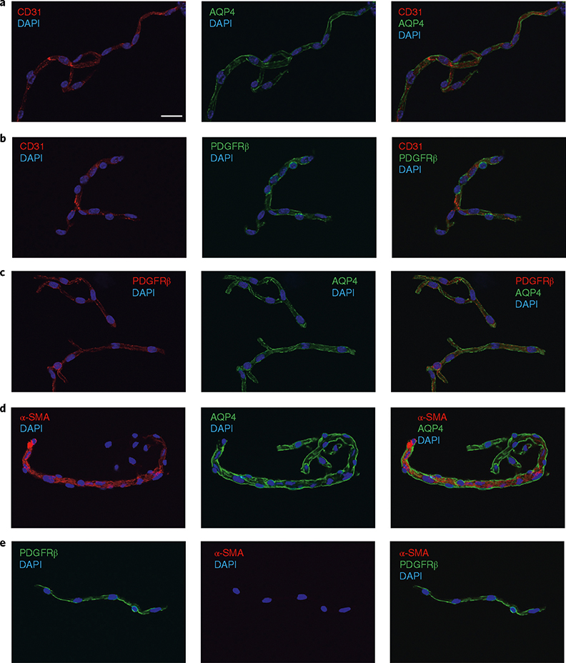
Fig. 3 |. Morphological characterization of microvessel preparations: structural integrity and cell composition.
a–e, Representative pictures of immunofluorescence analysis of isolated microvessels (≤10 μm, ~93% of all microvessels) stained with antibodies for each cell component. Endothelial markers (CD31, red channel; a,b), astrocyte end-foot markers (AQP4, green channel; a,c) and pericyte/smooth muscle cell markers (PDGFRβ, green or red channel; b,c) were detected in all microvessel fragments. d, Notice the transition of a precapillary arteriole (α-SMA+) to capillaries (α-SMA−) in the microvessel fragment. e, Example of an α-SMA− microvessel. DAPI (nuclear stain, blue channel). Images were taken using confocal laser scanning microscope (Olympus FV10i) and processed using Olympus Fluoview v.3.0. Scale bar, 20 μm.

