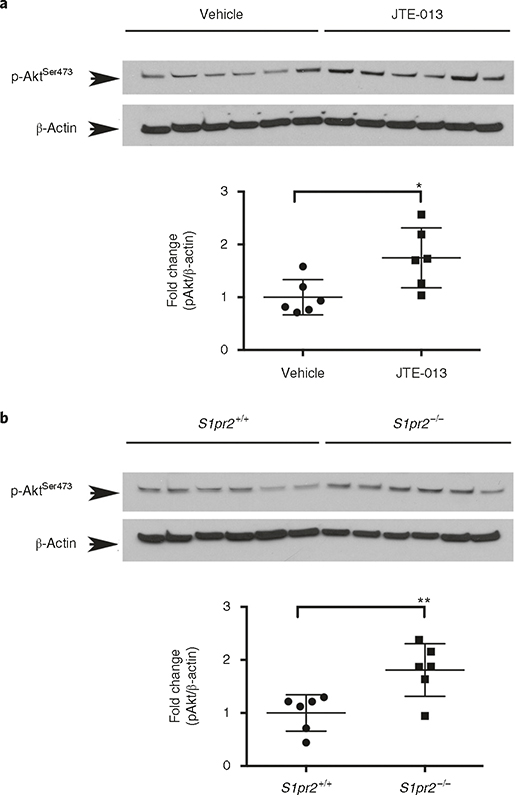
Fig. 7 |. Quantification of phospho-Ser473 Akt levels in cerebral microvessels in wild-type mice after administration of the S1PR2 antagonist JTE-013.
a, Mice were treated with vehicle or JTE013 (30 mg/kg) by gavage. Six hours later, microvessels were isolated and proteins were subjected to western blot analysis. b, Microvessels were isolated from 8-week-old adult S1pr2 knockout (S1pr2−/−) mice or wild-type littermates (S1pr2+/+), and western blot analysis for phospho-Ser473 Akt (pAkt) was conducted; n = 6. Optical density of each lane was measured using Image J. Fold induction of p-Akt (normalized by β-actin) in JTE013-treated versus vehicle-treated mice (a) and S1pr2−/− versus wild-type mice (b) are shown. The individual values and the average ± s.e.m. are shown. *P < 0.05, **P < 0.005 (t-test). The use of laboratory animals was approved by the Weill Cornell Medicine Institutional Animal Care and Use Committee. Full western blots for a and b are shown in Supplementary Fig. 1.

