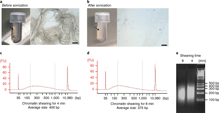
Fig. 9 |. Optimization of chromatin extraction and shearing from cortical microvessels.
Isolated microvessels were cross-linked with 1% (wt/vol) methanol-free paraformaldehyde and subjected to lysis and chromatin extraction protocol. a,b, Optimization of microvessel disruption and lysis. a, Images of lysate after vortexing for 15 min in lysis buffer B, per the manufacturer’s instructions. (Left) Arrow indicates visible microvessels in the microTUBE. (Right) Microscopic image of the lysate taken under inverted microscope (Olympus CKX41; scale bar, 100 μm). b), Images of lysate after optimization of the conditions to efficiently disrupt microvessels. (Left) Microvessels are no longer visible. (Right) Microscopic image of the lysate. c–e, Optimization of chromatin shearing. Impact of the time of sonication on the size of chromatin fragments. After microvessel disruption, extracted chromatin was sonicated for 4 min (c,e) or 8 min (d,e). The size of the sheared chromatin was determined with a Bioanalyzer (c,d) and electrophoresis on a 2% (wt/vol) agarose gel (e). Right lane: 100-bp DNA ladder. FU, fluorescence units.

