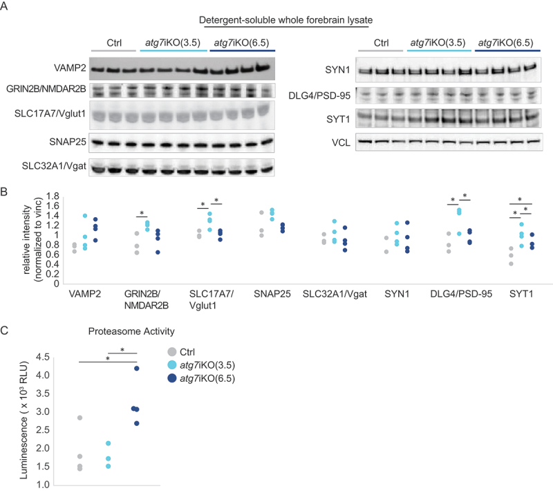
Figure 2.

Analyses of whole tissue homogenates suggests that cells compensate for autophagy deactivation over time. (A and B) Quantitative western blot analyses of detergent soluble lysates of mouse forebrain. Analyses reveal a significant accumulation of synaptic proteins GRIN2B/NMDAR2B, SLC17A7/Vglut1, DLG4/PSD-95, and SYT1 in atg7iKO(3.5) mice, which in atg7iKO(6.5) mice, return to Ctrl levels. VAMP2, SLC32A1/Vgat and SYN1, did not show any change. VCL was used as a loading control. Ctrl, n = 3; atg7iKO(3.5), n = 4; atg7iKO(6.5), n = 4. (C) Chymotrypsin-like activity of the proteasome in forebrain tissue of mice. atg7iKO(6.5) mice demonstrate a significant increase in proteasome activity. Ctrl, n = 3; atg7iKO(3.5), n = 4; atg7iKO(6.5), n = 4. Statistical analyses are found in Table S1. *p < 0.05.
