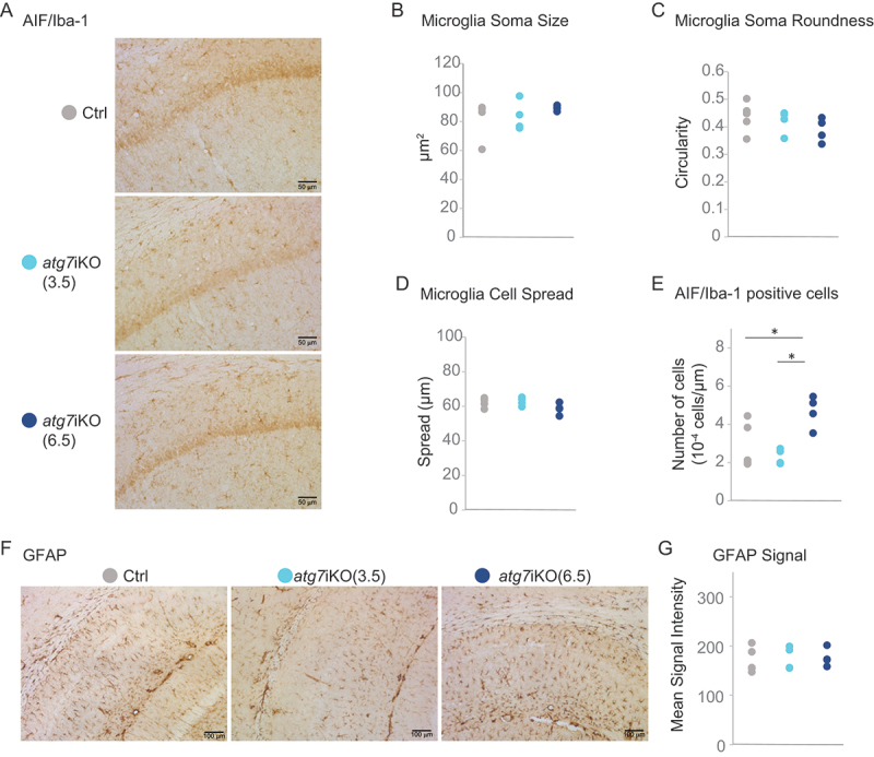
Figure 5.

Deactivation of autophagy does not result in neuroinflammation in the hippocampus. (A-E) Immunohistochemistry of coronal brain sections of hippocampus stained for AIF1/Iba1 as a marker for microglia. AIF1+ cells were assessed for (B) soma size, (C) soma roundness, (D) cell spread and (E) the total cell number. Scale bar: 50 µm (F and G) Staining against GFAP as a marker for astrocytes (G) Quantification for GFAP signal intensity. Scale bar: 100 µm. Ctrl, n = 5–6; atg7iKO (3.5 wks), n = 4; atg7iKO (6.5 wks), n = 4. Statistical analyses are found in Table S1. *p < 0.05.
