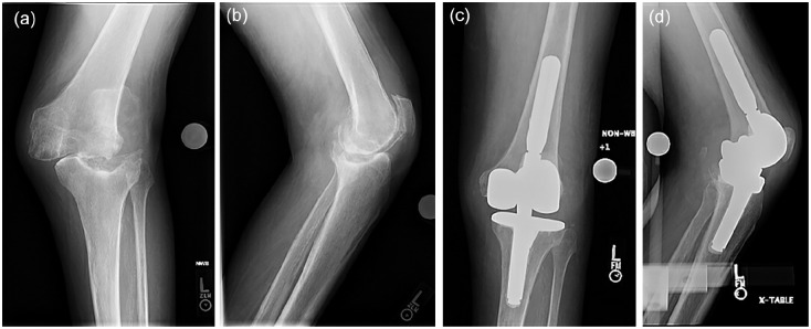
Fig. 2.
A 73-year-old patient with a history of idiopathic demyelinating neuropathy presented with left knee pain and varus deformity that progressed significantly over a year. He could not bear weight on his left leg due to knee pain and instability. Anteroposterior (a) and lateral (b) preoperative radiographs demonstrate lateral femoral-tibial joint subluxation, large central tibial bone defect, and bone fragmentation of the medial femoral condyle. The patient was indicated for primary TKA with a hinge implant. Intraoperatively, the medial collateral ligament was incompetent, and the tibial defect was filled with bone autograft. Postoperative anteroposterior (c) and lateral (d) radiographs demonstrated hinge implants with excellent alignment and no complication.

