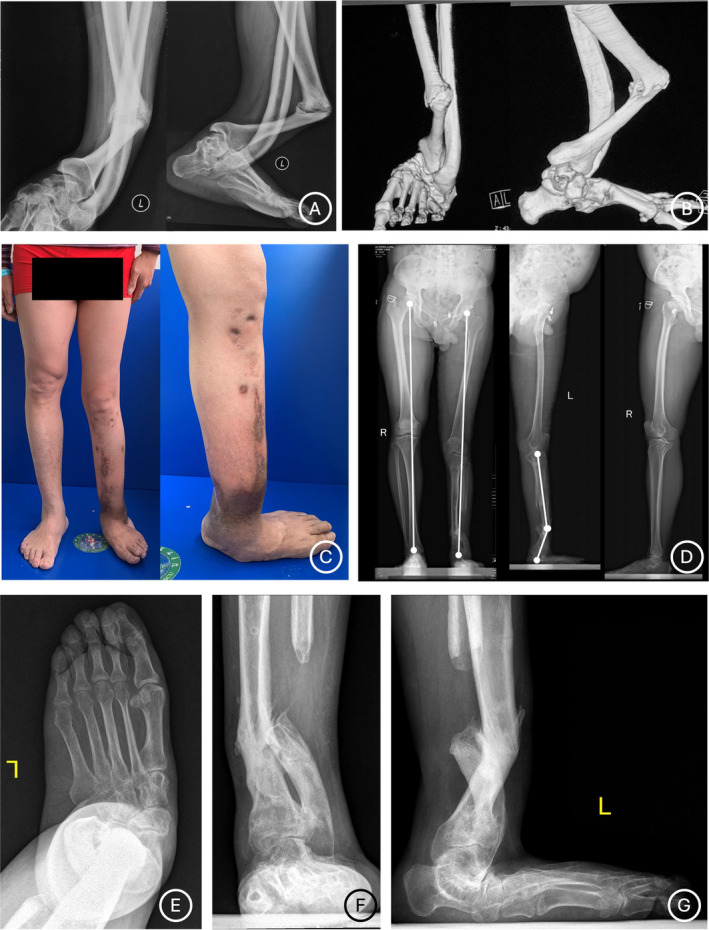
FIGURE 2.

Preoperatively clinical and radiographical photographs of a 58‐year‐old female patient (Case No. 10) who has a CPT in left lower limb and suffered from two previously failed surgeries. (A–B) Radiographical photographs before his previous surgeries show a CPT characterized with significant anterolateral bowing and varus deformities. (C‐G) Appearance and radiological images before our Ilizarov distraction show poor soft‐tissue condition and anterolateral bowing and valgus deformities at the CPT site. Serious secondary deformities of the ipsilateral foot characterized with a collapsed foot arch, valgus, and abduction are noted.
