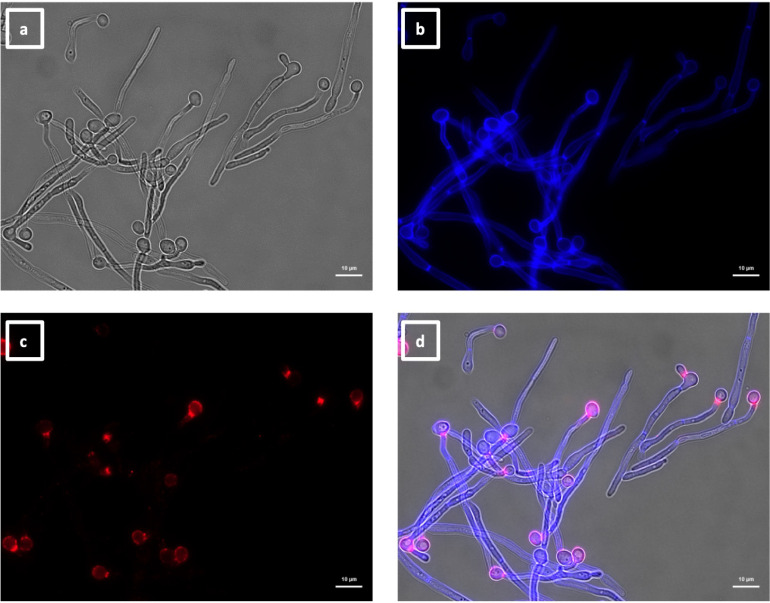
Fig 7.
Immunolabeling of C. albicans hyphae with VHH19. Hyphae of C. albicans strain SC5314 were grown in RPMI-1640 medium at 37°C for 6 h and fixed with formaldehyde. The samples were stained with calcofluor white and labeled with VHH19, and a rabbit anti-VHH polyclonal antibody and a goat anti-Rabbit IgG–Alexa Fluor 647–conjugated antibody for detection. Panels indicate: (a) DIC, (b) calcofluor white, (c) Alexa Fluor 647, and (d) merged view. Cells were imaged using a Zeiss Axio Imager M2 microscope.

