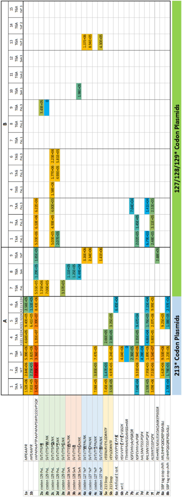
FIGURE 4.

Overall summary of termination codon readthrough or variant derived peptides in all replicates. The data summarized are from the same experimental data as in Figure 3 except the data contain peptide sequences that do not match wild-type p53. (A) Transient transfection. The 213-TAA p53 plasmids are presented in Column 1; the 213-TAG plasmid expression peptides are Column 2; and the 213-TGA plasmid expressing peptides are in Column 3. Three different p53 expression plasmids contained the three different termination codons of p53 at position 213 except these also contained two E-K mutations (221-EPPE-224; 221-KPPK-224) in order to introduce tryptic cleavage sites near the 213 termination codon in attempts to recover more tryptic peptides from this region; the 213-TAA plasmid peptides are presented in Column 4; the 213-TAG plasmid expression peptides are Column 5; and the 213-TGA plasmid expressing peptides are in Column 6. A third experiment used three different plasmids with the same TGA mutation at codons 127, 128, and 129 (columns 7–9). (B). Stably expressing p53 cells. As in Figure 8, the same samples are present; (i) the codon 129 (PxL) expression plasmids in triplicates (columns 1–3, TAA; columns 4–6, TAG; and columns 7–9, TGA); (ii) the codon 128 (SxA) expression plasmids in triplicate (columns 10–12, TGA); (iii) the codon 127 (YxP) expression plasmids in triplicate (columns 13–15, TGA). The numbers in each box represent ion intensity.
