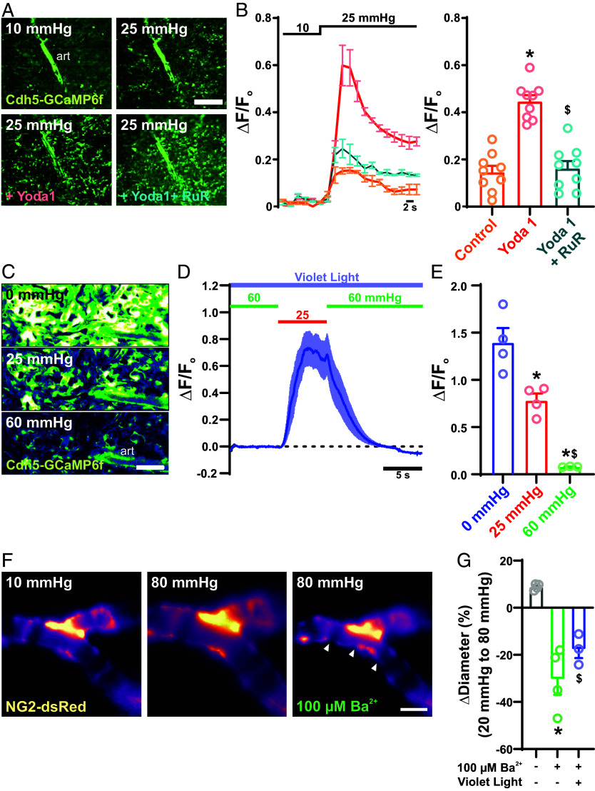
Fig. 2.
Intraluminal flow and pressure regulate light-dependent signaling. (A) Representative images of a choroidal preparation from a Cdh5-GCaMP6f mouse showing Ca2+ fluorescence in the choriocapillaris endothelium in response to increases in intraluminal pressure from 10 mmHg to 25 mmHg under control conditions and in the presence of bath-applied Yoda 1 (10 μM) or Yoda 1 + ruthenium red (30 μM) (n = 3 choroid preparations from 3 mice). (B) Traces (Left) and summary data (Right) showing averaged fluorescence of Ca2+ transients in the choriocapillaris endothelium in response application of Yoda 1 (10 μM) (error bars; *P < 0.05 to control and $P < 0.05 to yoda1. n = 3 choroid preparations from 3 mice per group). (Scale bars, 300 μm.) (C–E) Representative images (C), traces (D), and summary data (E) of Ca2+ transients in the choriocapillaris endothelium in response to violet light stimulation (405 nm, 6.1 × 10¹⁴ photons/cm2/s) at 0, 20, and 60 mmHg (error bars; *P < 0.05 to 0 mmHg and $P < 0.05 to 25 mmHg, n = 3 choroid preparations from 3 mice). (F and G) Representative images (F) and summary data (G) showing pressure-induced arteriole constriction in the absence of light stimulation, without and with the Kir channel blocker Ba2+ (100 µM), and with violet light stimulation (6.1 × 10¹⁴ photons/cm2/s) in the presence of Ba2+ (error bars; *P < 0.05 to control and $P < 0.05 to 100 µM Ba2+, n = 6 ROIs in 3 choroids from 3 mice per group).

