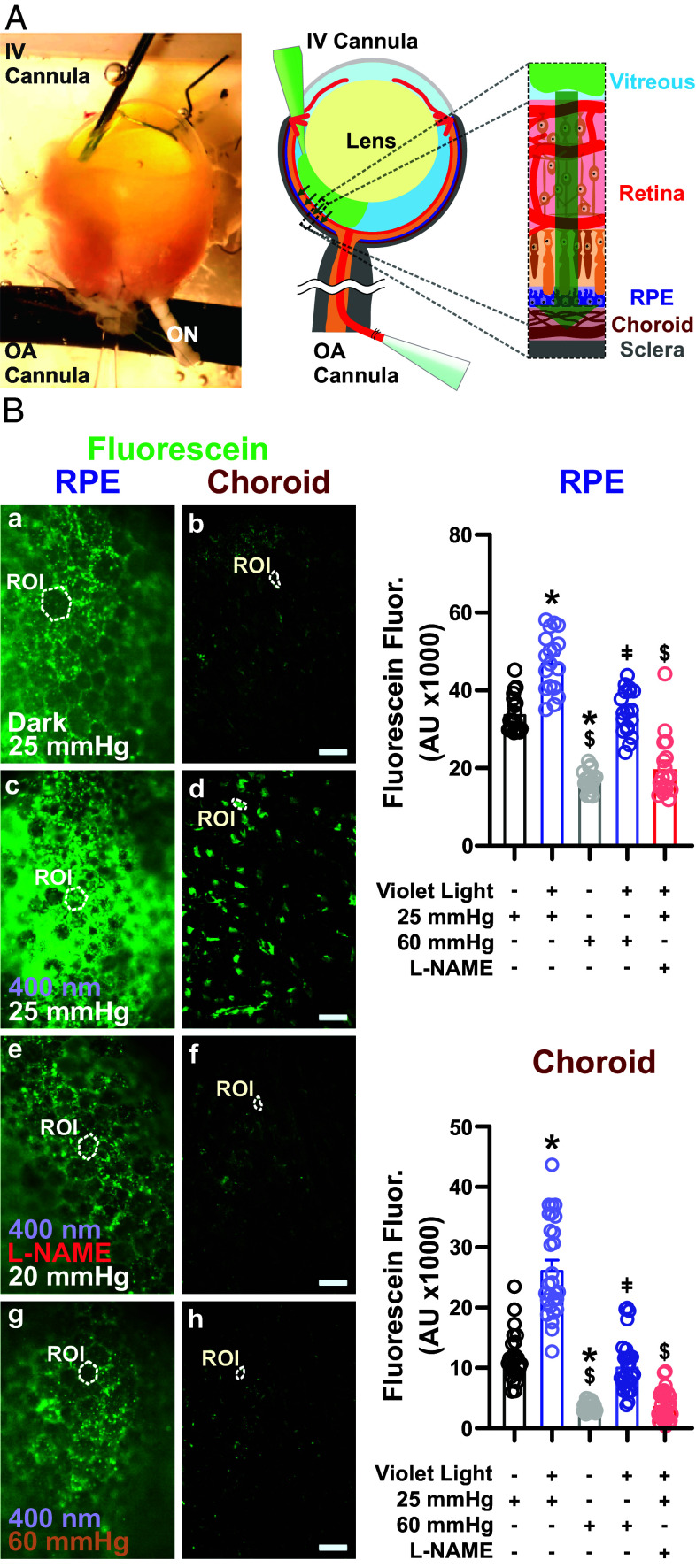
Fig. 3.
Photomechanical control of subretinal fluid absorption. (A, Left) Depiction of our ex vivo double-cannulation technique. The enucleated mouse eye is arterially cannulated using a glass pipette, and the vitreous chamber is cannulated using a fine needle for fluorescein injection (3 μM). (Scale bar, 500 µm.) (A, Right) Schematic illustration of intravitreal injection of sodium fluorescein and its absorption posteriorly into the choroid. (B, Left) Representative fluorescence micrographs showing fluorescein absorption into the RPE (Left) and choroid tissues (Right) from the vitreous space under dark conditions at an intraluminal pressure of 25 mmHg (a and b), with violet light stimulation for 60 min at an intraluminal pressure of 25 mmHg (c and d), with violet light stimulation (60 min) and intraluminal L-NAME (10 μM) infusion at 25 mmHg (e and f), and with violet light stimulation (60 min) at 60 mmHg. (Scale bars, 20 μm.) (B, Right) Summary data showing fluorescein signals in the RPE (Top) and choroid (Bottom) under the conditions indicated in B. Data are presented as means ± SEM (error bars; *P < 0.05 to dark treatment at 25 mmHg; $P < 0.05 to violet light treatment at 25 mmHg; and ǂP < 0.05 to dark treatment at 60 mmHg; n = 18 ROIs in 4 choroids from 3 mice per group).

