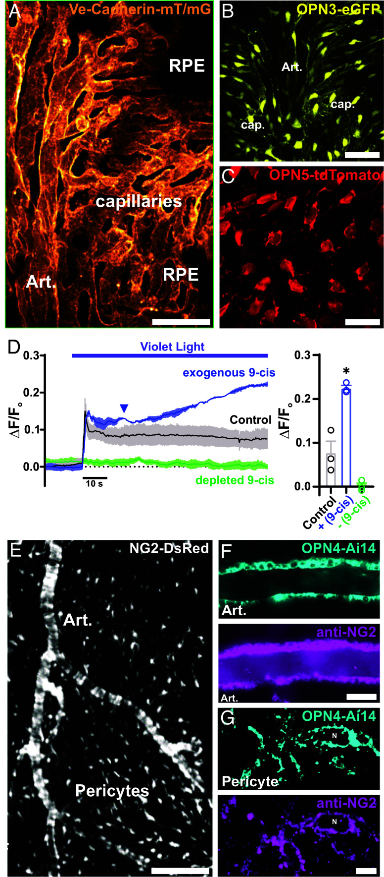
Fig. 4.
Expression of OPN3, OPN4, and OPN5 photopigments in the choroid vasculature. (A) Representative image displaying the ultrastructure of the choriocapillaris endothelium in choroid flat mounts from a Ve-cadherin mT/mG mouse. (B) Representative image showing the expression of encephalopsin or panopsin (Opn3) in choroid flat mounts from an Opn3-eGFP mouse. (Scale bar, 50 μm.) (C) Representative image showing the expression of neuropsin (Opn5) in choroid flat mounts from an Opn5-tdTomato mouse. (Scale bar, 25 μm.) (D) Representative trace and summary data of averaged GcAMP6f fluorescence in the choriocapillaris endothelium in response to constant violet light stimulation (405 nm, 6.1 × 10¹⁴ photons/cm2/s) under control conditions (gray), in the presence of exogenous 9-cis retinal (blue), and following depletion of 9-cis retinal (green). Data are presented as means ± SEM (error bars; *P < 0.05; n = 3 ROIs in 3 choroids from 3 mice per group). (E) Representative image showing the zonation of mural cells in choroid flat mounts from an NG2-dsRed mouse. (Scale bar, 40 μm.) (F and G) Representative images showing melanopsin expression in choroidal SMCs (F) and choroidal pericytes (G) in choroid flat mounts from an Opn4-Ai14 mouse and immunohistochemical detection of NG2 using an anti-NG2 polyclonal antibody. [Scale bars, 60 μm (Left) and 5 µm (Right).]

