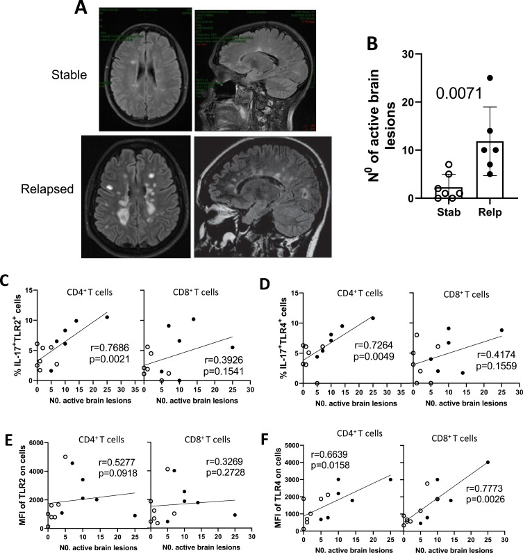
Figure 4.
The radiological activity of RRMS as a function of clinical activity of the disease and its relationship with TLR+ T cell subsets. The number of new active brain lesions, observed by MRI scans after 1 year of DMT, was determined in non-relapsed (Stab n=7) and relapsed (Relp n=6) RRMS patients during the 1-year follow-up. (A) Shows MRI scan of the brain in a control (above) and relapsed (below) RRMS patients showing typical active lesions. In (B), the mean ± SD number of brain lesions in Ctrl versus Relp (Student’s t test). From (C to F) the radiological activity was correlated with the percentage of IL-17-secreting (CD4+ and CD8+) T cells positive for TLR-2 (C) and TLR-4 (D), and MFI of TLR-2 (E) and TLR-4 (F) (Spearman correlation).

