Fig. 1.

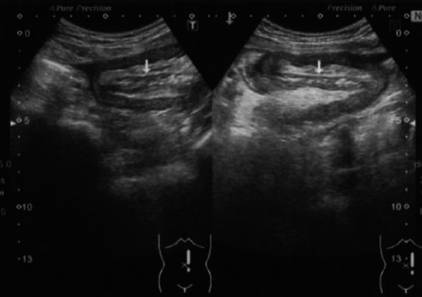
Abdominal ultrasonography. A small amount of ascites in the cysto-rectal fossa, a high echogenic mesentery, and small intestinal insertions in the intestinal tract of the left abdomen can be noted. Oral bowel dilatation was also observed

Abdominal ultrasonography. A small amount of ascites in the cysto-rectal fossa, a high echogenic mesentery, and small intestinal insertions in the intestinal tract of the left abdomen can be noted. Oral bowel dilatation was also observed