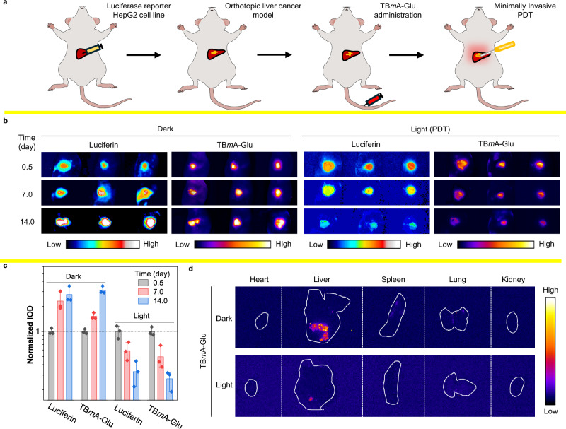
Fig. 7. Investigation of the therapeutic effect of TBmA-Glu mediated phototherapy in orthotopic liver cancer mouse models.
a Schematic diagram of the modeling process of a mouse model of liver tumor in situ. b The fluorescence images of the in situ hepatoma mice at the start, middle, and end of the treatment process by TBmA-Glu (5 mg/kg). Luminescence, λem = 550 ± 50 nm (Band-pass filter), 1000 ms. TBmA-Glu, λex = 500 nm, λem = 650 nm (Long-pass filter). c The normalized integrated optical density of cancer in the mice of in situ hepatoma models (n = 3) after treated TBmA-Glu (5 mg/kg) with or without light irradiation, data expressed as average ± standard error. d The fluorescence images of the separated organs of the orthotopic liver cancer mice at the end of the therapeutic process. TBmA-Glu, λex = 500 nm, λem = 650 nm (long-pass filter).

