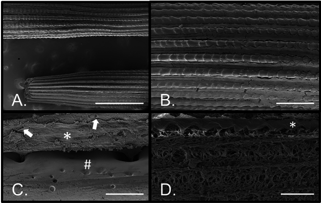
Fig. 6.
Cells of the spine revealed by scanning electron microscopy (SEM). (A) Low magnification view of two intact spines along their length showing the septa (mineralized ridges) and interseptal spaces (between the septa). (B) Higher magnification of a spine where left–right orientation is proximal-distal for the spine, respectively. Each of the septa have proximal-distal oriented ridges and are covered by the external epithelium. (C) The epithelium (*) covers the mineralized skeleton (#) with tightly adherent junctions (arrows) as well as projections through pores internally. (D) With most of the epithelium removed from the skeleton, except for the row identified by the asterisk (*), the cellular projections and extracellular matrix are apparent. Bar: A = 500 microns. B = 100 microns. C = 20 microns. D = 50 microns.

