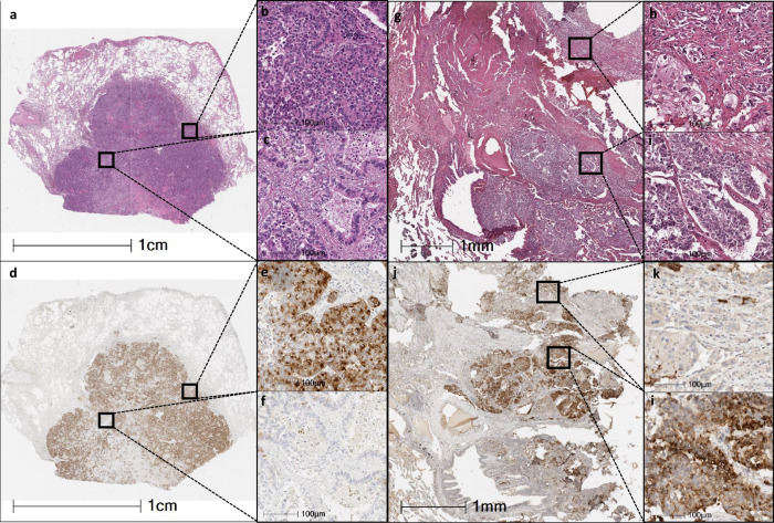
Fig. 4. Microphotographs of two combined tumors (large cell neuroendocrine carcinoma with adenocarcinoma).
Left side: low magnification microphotograph of combined tumor stained with H&E (×4). a Miniatures showing higher magnification of the large cell neuroendocrine carcinoma pattern (b) and adenocarcinoma acinar pattern (×40). c Low magnification of the same tumor stained with DLL3 (4X). d Miniatures of the same areas (b, c) showing large cell neuroendocrine carcinoma expressing DLL3 (e), and adenocarcinoma without DLL3 expression (×40) (f). Right side: Microphotograph at medium magnification (×10) of another combined tumor stained with H&E (g). Miniatures showing higher magnification of the large cell neuroendocrine carcinoma pattern (h) and adenocarcinoma solid pattern (i) (×40). Same area stained with DLL3 (j) (10X). Miniatures at higher magnification showing large cell neuroendocrine carcinoma expressing DLL3 (l) and adenocarcinoma without DLL3 expression (k) (×40).

