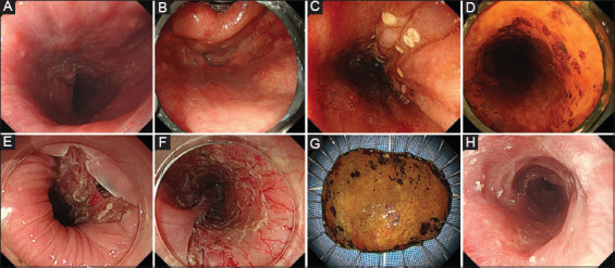
Figure 1.

Endoscopic submucosal dissection with pharyngeal expansion for cervical esophageal cancer. (A) The cervical esophageal cancer extends to the upper esophageal sphincter. The endoscope is unstable, and the endoscopic view is not clearly obtained because of the existence of the sphincter. (B) Pharyngeal expansion using a curved laryngoscope has maintained cervical esophageal broadening. (C) The oral margins of the lesion could be well-visualized and markings around the tumor are easily placed. (D) The lesion is expanded to three fourths of the circumference. (E) A mucosal incision is created at a sufficient distance from the oral edge of the tumor. (F) The resection area extends semi-circumferentially. Local injection and oral administration of steroids are provided. (G) The lesion is removed in an en bloc fashion with negative tumor margins. (H) No symptomatic stricture has developed 2 months later
