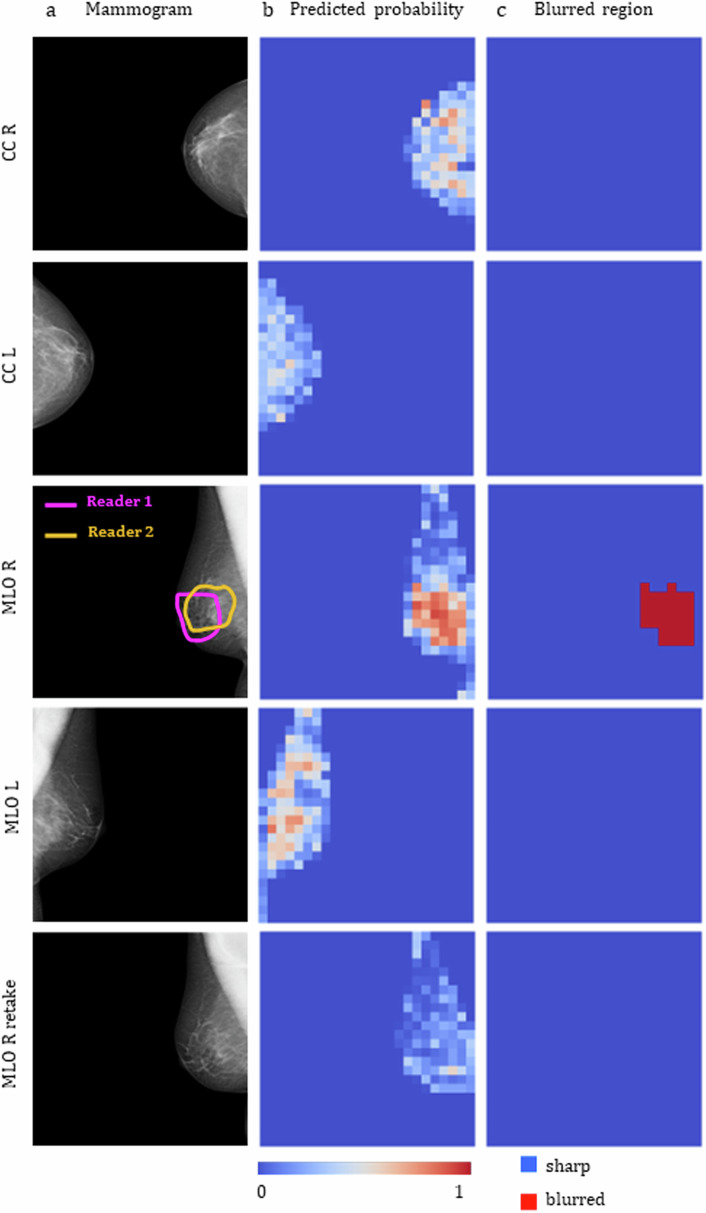
Fig. 6.
Blur detection results for a mammographic examination contained in the test set. a The right MLO mammogram in the original examination was labelled by both Readers 1 and 2 as blurred, the retake of this view is also shown in the bottom line. b Blur probability maps output by the trained model. c Final blurred area classification obtained after thresholding. CC, Craniocaudal view; L, Left breast; MLO, Mediolateral oblique view; R, Right breast

