. 2024 Nov 17;12(11):e009522. doi: 10.1136/jitc-2024-009522

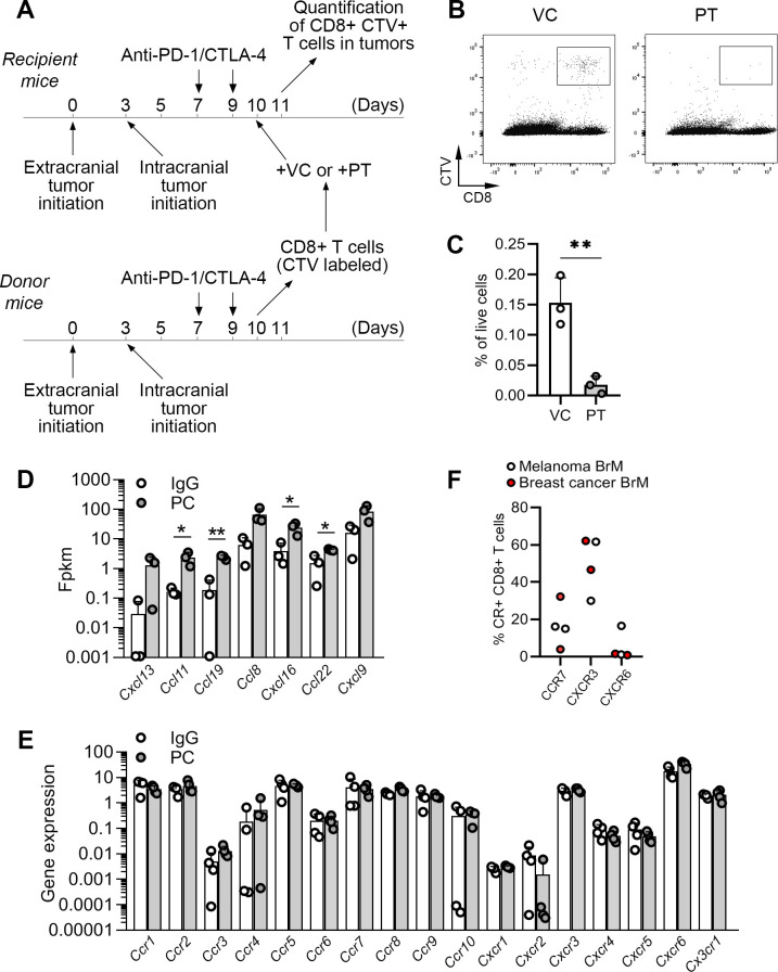
Figure 5. Trafficking of CD8+T cells into intracranial tumors is chemokine-dependent. (A) Experimental timeline for adoptive transfer of CD8+T cells. T cells isolated from donor mice were ex vivo labeled with CellTrace Violet (CTV) and treated either with pertussis toxin (PT) or vehicle control (VC) prior to their transfer into recipient mice. (B) Representative dot plots showing detection of adoptively transferred CTV+CD8+ T cells (following their ex vivo treatment with PT or VC) in intracranial tumors 18 hours post-transfer. (C) Quantification of data shown in B. (D) Analysis of chemokine gene expression in mRNA-seq data shown in figure 3. (E) Quantification (PCR) of chemokine receptor expression in CD8+T cells isolated from murine intracranial tumors by FACS. (F) Quantification of chemokine receptor expression on CD8+T cells within human brain metastases (n=2 for melanoma and n=2 for breast cancer) by flow cytometry. Statistical significances in C and D were determined by unpaired two-tailed t-test; *p≤0.05, **p≤0.01. BrM, brain metastases; CTLA-4, cytotoxic T-lymphocyte-associated protein 4; IgG, immunoglobulin G; mRNAseq, messenger RNA sequencing; PC, PD-1/CTLA-4; PD-1, programmed death-1; FACS, Fluorescence-activated cell sorting; Fpkm, Fragments Per Kilobase of transcript per Million mapped reads.

Figure 5