
Figure 1. CXCR2-deficient mice promote ear tissue regeneration.
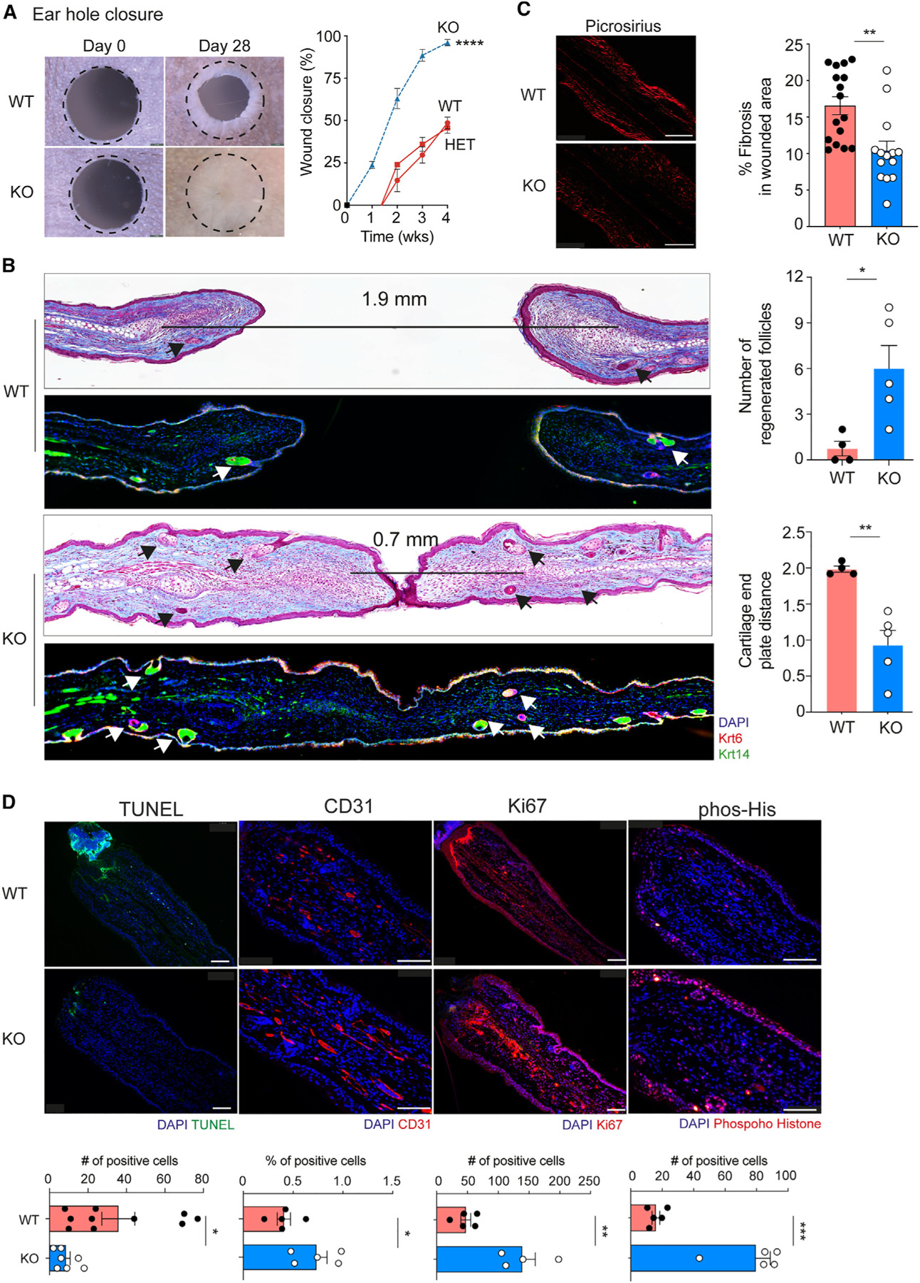
(A) Representative photographs and percentage of wound closure in wild-type (WT, n= 5), heterozygous (HET, n= 12), and CXCR2 KO homozygous (KO, dashed blue line, n = 6) mouse ears. A dotted circle represents the original 2-mm hole. 2-way ANOVA with KO compared to WT or heterozygous.
(B) Representative trichome stain and immunofluorescence of wounded ear tissue sections depicting neogenic hair follicles (Krt14+, Krt6+) from WT (n = 5–6) and CXCR2 KO (n = 5) mice. The distance between cartilage endplates is denoted by a black bar. Arrows indicated regenerated skin appendages. Right: quantitation of hair follicles in healed areas and distance between cartilage endplates. Unpaired two-tailed Student’s t test.
(C) Representative photographs and percentage of fibrosis assessed by picrosirius red staining in WT (n = 16) and KO (n = 14) wounds. Scale bars, 100 μM. Unpaired two-tailed Student’s t test.
(D) Representative images and quantification of immunofluorescence of tissue sections from wounded WT and CXCR2-KO mouse ears for apoptosis (TUNEL, n = 7), angiogenesis (CD31, n = 5) and cell proliferation (phosphorylated histone H3, n = 5 and Ki67, n=5 for WT, and n=4 for KO). Scale bars, 100 μM. Unpaired two-tailed Student’s t test. *p < 0.05, **p < 0.01, ***p < 0.01. Mean ± SEM are plotted.