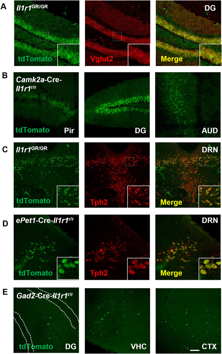
Fig. 2.
Neuronal IL-1R1 is expressed in diverse types of neurons classified by different neurotransmitters. (A) Representative images of tdTomato and Vglut2 labeling in Il1r1GR/GR hippocampal sections. Dashed squares mark the area shown at higher magnification on the bottom right. (B) Representative images of tdTomato from different CamK2a-Cre-Il1r1r/r brain sections. Pir, Piriform cortex; DG, dentate gyrus; AUD, primary auditory cortex. (C) Representative images of tdTomato and Tph2 labeling in Il1r1GR/GR dorsal raphe sections. A dashed square marks the area shown at higher magnification on the bottom right. DRN, dorsal raphe nucleus. (D) Representative images of tdTomato and Tph2 labeling in ePet1-Cre-Il1r1r/r dorsal raphe sections. Dashed squares mark the area shown at higher magnification on the bottom right. (E) Representative images of tdTomato from different Gad2-Cre-Il1r1r/r brain sections, white dotted lines denote granule cell layer of the DG. DG, Dentate gyrus, VHC, ventral hippocampus, CTX, cortex. Scale bar: 200 μm

