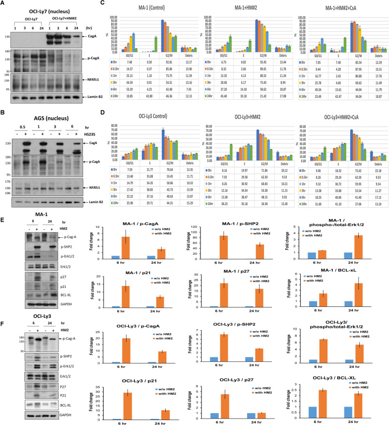
Fig. 2.
CagA, tyrosine-phosphorylated CagA, and NFATc1 can be translocated to the nucleus in HP-co-cultured GCB-DLBCL and gastric epithelial cells, CagA-related signaling molecules in HP-co-cultured DLBCL cell lines, and HP-co-cultured MA-1 cells and OCI-Ly3 cells exhibit G1 cell-cycle retardation. A In HP (HM#2)-co-cultured OCI-Ly7 cells, nuclear CagA expression was detected 1 and 3 h after HP stimulation; however, the effect decreased gradually at 6 h and decreased predominantly at 24 h. The expression of p-CagA in the nucleus was detected at 1 and 3 h; however, its signal intensity decreased at 6 h and eventually disappeared at 24 h. Simultaneously, the nuclear expression of NFATc1 was detected at 1, 3, 6, and 24 h after HP stimulation. B In HP (HS235)-co-cultured AGS cells, CagA and p-CagA translocated to the nucleus at 0.5 and 1 h. However, the signal intensity of both CagA and p-CagA decreased at 3 and 6 h. Simultaneously, nuclear expression of NFATc1 was detected at 0.5 and 1 h after HP stimulation; however, its signal intensity decreased at 3 and 6 h. C Compared with non-HP-co-cultured MA-1 cells, G1 arrest was predominantly detected at 24 h in HP (HM#2)-co-cultured MA-1 cells. However, G1 phase arrest was reversed after administration of CsA in HP (HM#2)-co-cultured MA-1 cells. The results were expressed in triplicate for each treatment group and measured by flow cytometry analysis (error bar means standard error). D Compared with that noted in non-HP-co-cultured OCI-Ly3 cells, G1 arrest was predominantly detected at 24 h in HP (HM#2)-co-cultured OCI-Ly3 cells. After the administration of CsA to HP (HM#2)-co-cultured OCI-Ly3 cells, the G1 arrest at 24 h was reversed. E Immunoblotting showed that HP induced the expression of CagA-related signaling molecules, including p-CagA, p-SHP2, p-ERK1/2, p21, p27, and Bcl-xL in HP (HM#2)-co-cultured MA-1 cells at 6 and 12 h. Quantification of western blotting in (E) showed that the expression levels of p-CagA, p-SHP2, p21, and p27 were higher at 6 than at 24 h, whereas levels of p-ERK1/2, and Bcl-xL were higher at 24 than at 6 h. F In HP (HM#2)-co-cultured OCI-Ly3 cells, HP provoked the expression of p-CagA, p-SHP2, p-ERK1/2, p21, p27, and Bcl-xL at 6 and at 12 h. Quantification of western blotting in (F) showed that the expression levels of p-CagA, p-SHP2, p-ERK1/2, p21, p27, and Bcl-xL were higher at 6 than at 24 h

