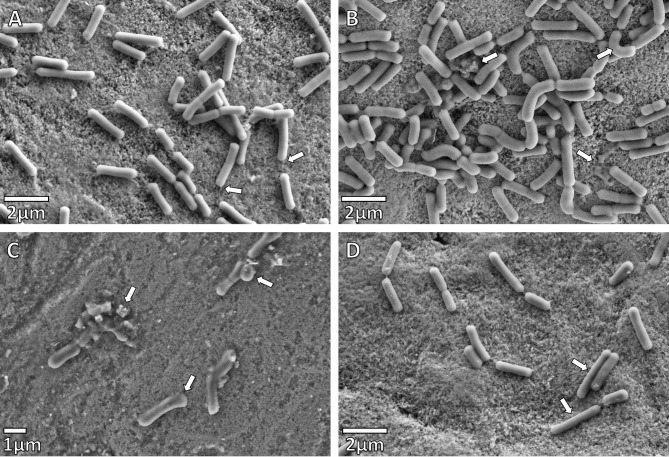
Fig. 4.
SEM micrographs of Lactobacillus paracasei after 48 h (A) Negative control. These rods are slow-growing bacteria. Adherence to the enamel surface observed with pellicle formation (indicated by arrows). (B) Enamel after Treatment 1: L. paracasei was added one hour after the synergistic plant combination at half-MIC. The normal morphology of L. paracasei, as can be seen in (A), has become distorted (upper right arrow). Cell debris is also indicated. (C) Enamel after Treatment 2: L. paracasei was added together with the synergistic plant combination at half-MIC. The normal morphology of L. paracasei is distorted giving the rods a club-like appearance, cells of different sizes and cell debris. (D) Enamel after Treatment 3: L. paracasei was added one hour before the synergistic plant combination at half-MIC. The normal morphology of L. paracasei, as can be seen in the 48-hour control sample (A) has become distorted with long thin cells now evident (indicated by arrows)

