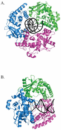
Figure 1.

The trimeric structure of λ Exo. View of the λ Exonuclease trimer looking through the central channel (A) and the same view rotated 90° to the right (B). The three subunits are colored blue, green, and magenta. The dsDNA passes through the central channel of the trimer, is acted upon by one of three active sites, and exits out the back as ssDNA. The structures were generated by PyMol based on the coordinates described by Zhang et al. (53).
