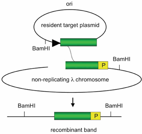
Figure 5.

Substrates used to demonstrate Red-promoted replisome invasion. Recombination occurs between a replicating resident target plasmid (direction of replication shown by black triangle) and a nonreplicating λ chromosome. The homologous regions are denoted by the green box. The λ chromosome is delivered at high efficiency by infection, is inhibited from replicating by overexpression of the λ c1 repressor, and is cut in vivo by a chromosomally encoded PaeR7 restriction enzyme. DNA from the infected cells is isolated at different times after infection, cut with BamHI, and subjected to a Southern procedure. The amount of recombinant band (bottom) is detected by probing a Southern blot for sequences designated “P.” (Descriptions of substrates were derived from reference 3).
