
Figure 6.
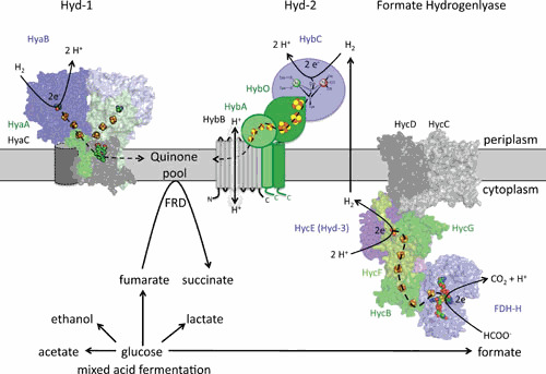
The hydrogenase-1 (Hyd-1), Hyd-2, and formate hydrogenlyase (FHL) complex in the cytoplasmic membrane of E. coli. The horizontal gray bar represents the cytoplasmic membrane. Components in each complex that have similar functions and exhibit amino acid sequence similarity share the same color. Large subunits are shown in blue tones, small subunits in greens, and integral membrane subunits in gray. The metal cofactors are shown as spheres with FeS clusters in brown/yellow, the [NiFe] cofactor in green/brown, and the molybdopterin guanine dinucleotide is shown as spheres in FDH-H. The Hyd-1 structure is based on PDB entry 4GD3 with one heme b molecule; an additional b-type cytochrome subunit has been added as a cylinder. A structure prediction based on complex I is shown for FDH-H and the Hyc components that form the FHL complex. Dashed arrows show the putative path of electron flow through each complex. The lower part of the panel shows the products of the mixed acid fermentation, of which succinate is generated by reduction of fumarate by fumarate reductase (FRD) using electrons derived from the quinone pool. The formate generated is the substrate for the FHL complex, yielding H2, which can be partially reoxidized by Hyd-1 and Hyd-2.