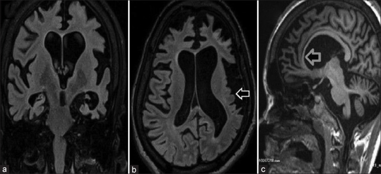
Figure 1.

Coronal (a) and axial (b) sections show atrophy of bilateral cerebral hemispheres, predominantly affecting the left cerebral hemisphere with microgyria involving the left parietal and temporal lobes (white arrow). There is asymmetric dilatation of both the lateral ventricles (left is larger than right) as seen in coronal (a) and axial (b) sections. Sagittal section (c) shows thinning of the corpus callosum (gray arrow)
