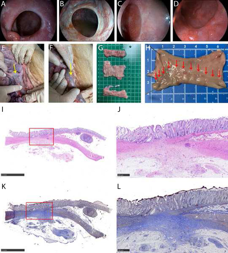
FIGURE 4.
Representative images of the anastomotic site. A, The anastomosis healed well on POD 30; endoscopy could be easily passed without intervention. B, Endoscopic appearance on POD 90. Small vessels extending radially from the anastomotic line and the white anastomotic edge were typical of what would be seen normally. C and D, Endoscopic appearance on POD 180 and POD 360 made it difficult to identify the anastomosis with only a faint line typically seen. E and F, The serosal aspect of a well-healed colonic anastomotic site (yellow arrow). G, The anastomotic scar and 6 cm above and below the anastomotic line were evaluated. H, Mucosal aspect of a well-healed colonic anastomotic site (red arrow). I and J, HE staining of the anastomosis site: the healing condition was good, as evidenced by a complete colonic mucosa with a mature morphology, fibrous tissue hyperplasia, and collagen deposition. Muscle bundles in the tunica muscularis are disorganized and partially replaced by collagen, fusing with the muscularis mucosa. K and L, Masson trichrome staining of the anastomosis site: hyperplastic fibrous tissue hyperplasia and collagen are green-blue, whereas hyperplastic smooth muscle cells are red. HE = hematoxylin-eosin; POD = postoperative day.

