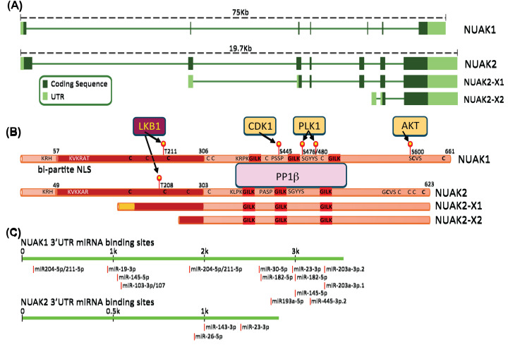
Figure 1. Structural alignment of NUAK1 and NUAK2.
(A) Schematic of primary transcripts encoded by NUAK1 and NUAK2 including introns and UTRs. (B) Alignment of NUAK1 and NUAK2 proteins. Experimentally validated phosphosites and upstream kinases are indicated, along with the positions of GILK motifs, nuclear localisation sequences, and approximate location of cysteine residues (conserved cysteines in bold print). Alignment of putative NUAK2-X1 and NUAK2-X2 isoforms is shown. (C) Alignment of miRNAs targeting the NUAK1 or NUAK2 3′UTR.

