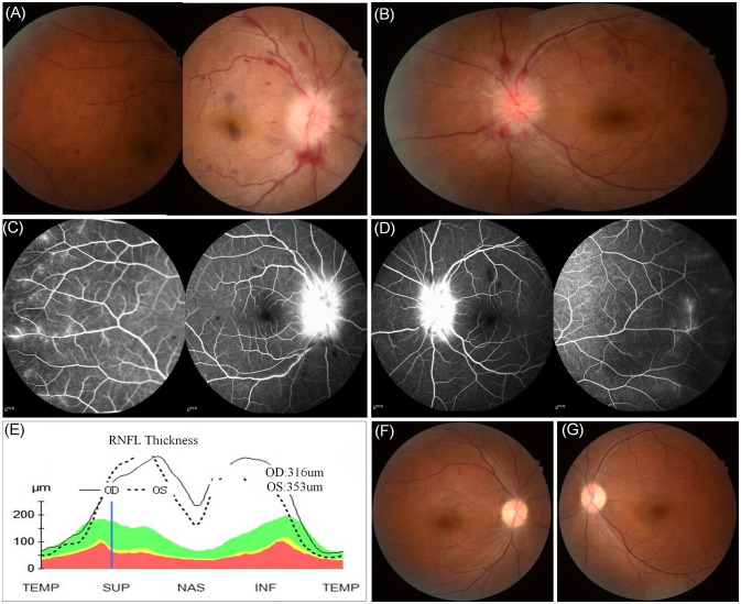
Figure 1.
Fundus examination revealed the presence of binocular peripapillary and perivascular haemorrhages as well as extensive deep retinal haemorrhages with severe optic disc swelling greater in the right eye (A) than in the left (B). (C, D) Fluorescein angiography images showed leakages of the bilateral optic disc and peripheral capillaries. (E) Retinal nerve fiber layer thickening on optical coherence tomography. (F, G) Retinal photos 1 month after onset showing the retinal haemorrhages in the binocular region had been absorbed completely.

