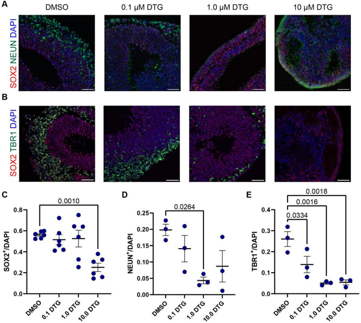
FIGURE 3.
Exposure to DTG reduces the number of postmitotic neurons in organoids. (A,B) Representative images of selected regions in Day 40 organoids from the C1-2 iPSC line following daily exposure to DTG from Day 7 to Day 39. Scale bars, 100 μm. (A) Expression of NEUN, a marker expressed in most postmitotic cortical neurons. (B) Expression of TBR1, a transcription factor expressed in postmitotic neurons during glutamatergic neuron development and SOX2, a marker of neural stem cells. Quantifications show a reduction in the proportion of SOX2+ neural stem cells (C), and postmitotic neurons expressing NEUN (D) or TBR1 (E) among DAPI cells under some conditions. Data points represent marker expression quantified from a single organoid region, with n = 3 organoid regions for TBR1 and NEUN expression and n = 6 for SOX2 expression. One-way ANOVA with Dunnett’s test for multiple comparisons, adjusted p values shown for statistically significant differences among pairwise comparisons. Data shown are means ± SEM.

