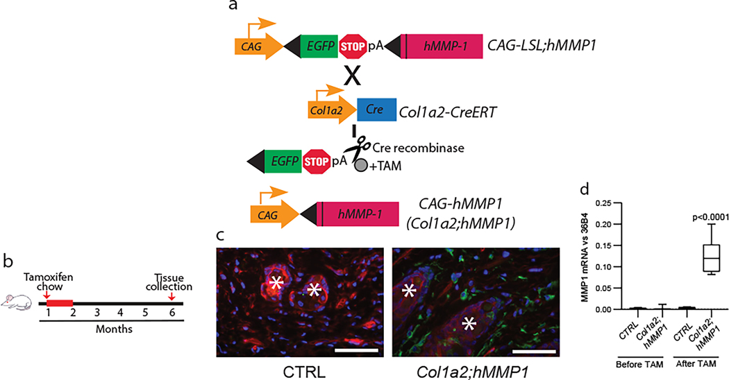
Figure 1. Generation of a humanized mouse model of dermal aging by fibroblast-selective expression of hMMP1. MMP1.
(a) Schematic representation of the generation of a mouse model of fibroblast expression of full-length, catalytically-active human MMP1 (hMMP1, Col1a2;hMMP1 mice). hMMP1 expression is activated by tamoxifen (TAM)-inducible Cre recombinase under the control of the fibroblast-specific Col-1a2 promoter and upstream enhancer. (b) Schematic diagrams depicting the initial feeding of one-month-old mice with TAM-containing chow (400mg/kg) for one month and back skin samples were collected at six months of age for analyses. (c) Cre recombinase activity (GFP) in the dermal fibroblasts of Col1a2-Cre(ER)T;ROSA26mTmG reporter mice. One-month-old mice were fed TAM chow for one month and the back skin was harvested at six months of age. The mTmG transgene confers expression of membrane-targeted tandem timer (td)Tomato prior to Cre-mediated excision and membrane-targeted green fluorescent protein (GFP) after excision. Tissue was counterstained with DAPI to allow visualization of nuclei. Representative images are shown. *Hair follicles. Bar=100 μm. (d) hMMP1 transgene mRNA expression in Col1a2;hMMP1 mice. The back skin samples were collected from one-month-old mice without tamoxifen administration (Before TAM) or six months after tamoxifen administration (After TAM). hMMP1 mRNA levels were determined by RT-PCR (normalized to 36B4, housekeeping gene internal control). N=5 for each group. Statistical analyses (t-test) were performed to evaluate the significance between the two groups. All P-values are considered significant when <0.05.

