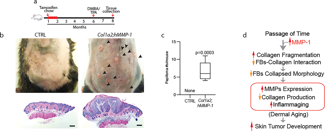
Figure 4. MMP1-mediated dermal aging promotes skin papilloma formation.
(a) Schematic diagrams depicting the initial feeding of one-month-old mice with TAM-containing chow (400mg/kg) and two-stage carcinogenesis. Back skin from Col1a2;hMMP1 and littermate hMMP1 negative control (six months after TAM treatment) mice were treated with a single dose of DMBA (100μg/100μl acetone) followed by biweekly applications of TPA (25μg/100μl acetone) for 8 weeks (two-stage chemical carcinogenesis). (b) Representative images of mice from each group are shown. Multiple skin tumors (black arrowheads) are seen in Col1a2;hMMP1 mice (upper right panel), while no tumors were observed in the control mice (upper left panel). Representative histology of treated back skin from control (lower left panel) and Col1a2;hMMP1 (lower right panel) mice. Note severe epidermal papillomatous dysplasia in Col1a2;hMMP1 mice. Bars=200μm (c) Quantitation of tumor numbers in control and Col1a2;hMMP1, following chemical carcinogenesis treatment. N=5 for each group. Statistical analyses (t-tests) were performed to evaluate the significance between the two groups. The P-value is considered significant when <0.05. (d) Proposed model for MMP1 mediated dermal aging and skin tumor formation: Age-related elevation of MMP1 activity degrades collagen fibrils thereby disrupting fibroblast-collagen interactions, resulting in reduced spreading and adaptive functional alterations that perpetuate further dermal ECM fragmentation (dermal aging) and creates a dermal microenvironment that is conducive to skin tumor development.

