FIGURE 1.

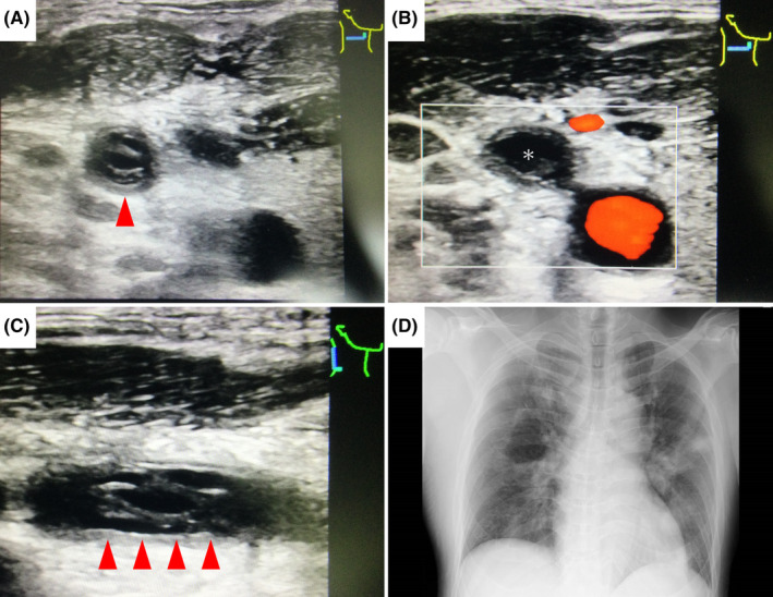
(A–C) Ultrasound image shows a hyperechoic thrombus in the right internal jugular vein (red arrowhead), and color Doppler ultrasound image demonstrates absence of flow (*). (D) Chest X‐ray shows multiple consolidations in both lungs.

(A–C) Ultrasound image shows a hyperechoic thrombus in the right internal jugular vein (red arrowhead), and color Doppler ultrasound image demonstrates absence of flow (*). (D) Chest X‐ray shows multiple consolidations in both lungs.